Project 1: Metastatic Initiation by Tumor Derived Extracellular Vesicles
Tumor Derived Exosome Induced Reprogramming to promote metastatses. A) Primary Tumor Cell in mouse mammary tissue (inset) secreted exosome. B) Exosome engulfment of stromal cells to initiate cell reprogramming. C) reprogrammed cells induced metastatic state & D) Metastatic cascade.
Extracellular vesicles (EVs) are small membrane-bound particles that are released by cells. They can contain a variety of cargo, including proteins, lipids, nucleic acids, and metabolites. EVs have been shown to play a role in a number of cellular processes, including tumor invasion. Exosomes, oncosomes and micro-vesicles are types of EVs studied in the lab. Exosomes are a type of extracellular vesicle that are used by normal and tumor cells to promote
Primary breast tumors exploit EVs as mediators of intercellular communication to promote cellular invasion and facilitate the metastatic spread of cancer. We are investigating the mechanisms used by tumor derived EVs to the development of novel therapeutic strategies to prevent or inhibit breast cancer metastasis.
We are exploring various ways that primary tumors use EVs to facilitate cellular invasion:
EV delivery of proteins like MMP13 to recipient cells can that promote tumor cell motility and invasion via breakdown extracellular matrix to initiate cell invasion.
EV delivery of microRNAs (miRNAs) that alter the gene expression of recipient cells to facilitate cellular reprogramming, modifying the microenvironment to initiate breast cancer metastasis to the bone.
Relevant Publications:
Li, G., Chen, T., Dahlman, J., Eniola-Adefeso, L., Ghiran, I. C., Kurre, P., Walker, ND,.. & Sundd, P. Current challenges and future directions for engineering extracellular vesicles for heart, lung, blood and sleep diseases. Journal of extracellular vesicles, (2023) 12(2), 12305.
White, ED, Walker, ND, Yi, H., Dinner, AR, Scherer, NF, Rosner, MR,. (2022) Volumetric microscopy of CD9 and CD63 reveals distinct subpopulations and novel structures of extracellular vesicles in situ in triple negative breast cancer cells. bioRxiv. doi: https://doi.org/10.1101/2022.11.08.515679.
Rabe DC, Walker, ND, Rustandy FD, Lee Jiyoung and Rosner, MR., Tumor Extracellular Vesicles Regulate Macrophage Driven Metastasis through CCL5. Cancers (Basel). 2021 July 10;13(14):3459. doi:10.3390.
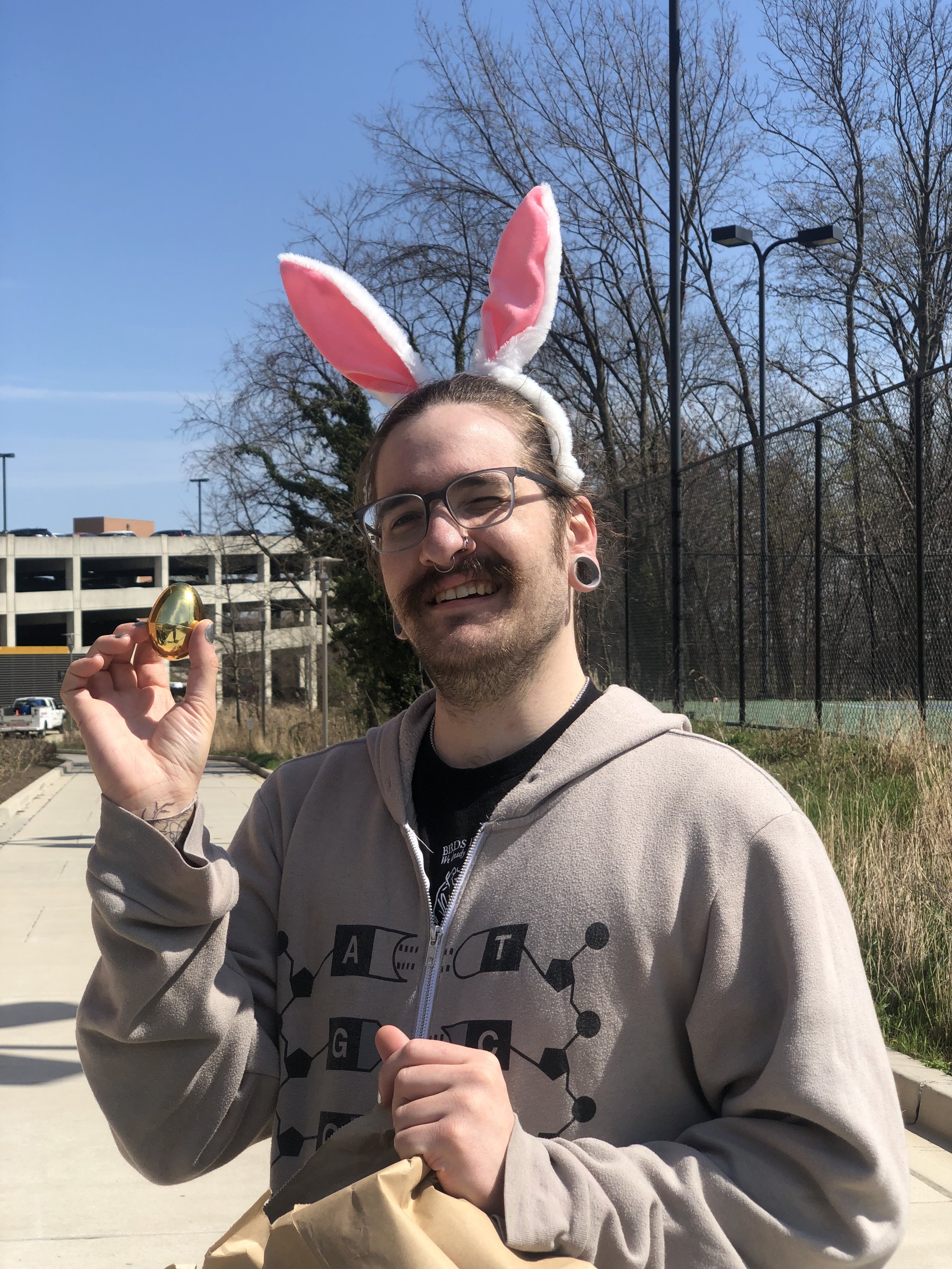
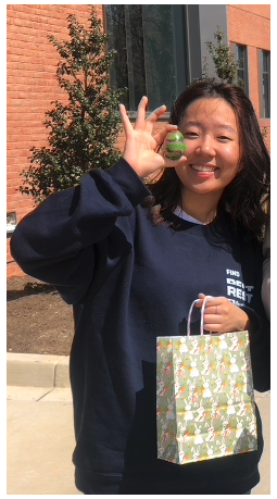
The team members involved are called the “INHIBITORS”, who are on a quest to discover mechanisms used by primary breast tumors to initiate metastasis wthat could serve as potential therapeutic strategies or early diagnostic biomarkers. Annually, we have a lab retreat that ends with an Egg-o-some hunt filled with candy cargo (see lab actives for details).
Meet the Inhibitors
-

Nykia Walker, PhD
Principle Investigator
The lab studies how different types of extracellular vesicles regulate transcriptional changes to initiate breast cancer metastasis. We use bioinformatics, molecular biology, gene editing tools, and proteomics to study transcriptional regulation .
-

Tyler Offenbacher
Master Inhibitor
Tyler is working to understand how exosome cargo, like microRNA regulates transcriptional expression of stromal cells in the tumor microenvironment and in distant stroma.
-

Joanna Chang
Oncosome Operative
Joanna is developing quantitative tools to measure large oncosomes in TNBC xenograft murine mammary tissue.
-

The Original Inhibitors
Ayo Oluwafemi & Madi Kore are alumni who started the histology and morphology studies of early breast cancer metastases diagnostic markers.
-

Are you the one?
We are recruiting new PhD students to explore how DEGs are controlled by tumor-derived exosomes and how they enable cell invasion.
